41 colon diagram with labels
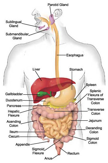
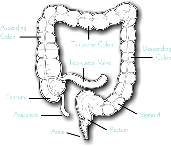
Large intestine with labels for the appendix, cecum ... Large intestine with labels for the appendix, cecum, ascending colon, transverse colon, descending colon, sigmoid colon, rectum, and anus View full-sized image Download Media Please credit each image as: National Institute of Diabetes and Digestive and Kidney Diseases, National Institutes of Health. Colon (Large Intestine): Anatomy, Function, Structure Sigmoid colon: The S-shaped connection between the last part of the colon and the rectum, located on the bottom left side of the abdomen is called the sigmoid colon. 2 Size and Length This organ is called the large intestine because of the diameter (width) of the intestine; it is much wider than the small intestine, but also much shorter.
Picture of the Human Colon Anatomy & Common Colon ... - WebMD The ileum (last part of the small intestine) connects to the cecum (first part of the colon) in the lower right abdomen. The rest of the colon is divided into four parts: • The ascending colon...

Colon diagram with labels
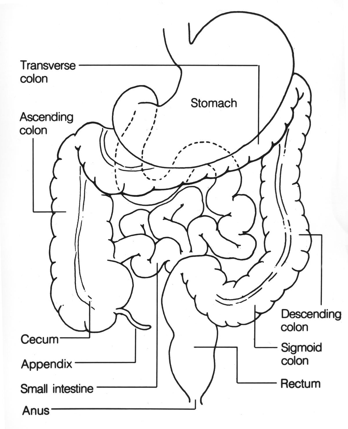
Anatomy of the Human Pancreas Explained With Labeled Diagrams Anatomy of the Human Pancreas Explained With Labeled Diagrams. The pancreas, that somewhat look like the head of a golf club, does the job of producing digestive juices. It can be divided into 4 parts―the head, neck, body, and the tail. Bodytomy elaborates more on the anatomy of the human pancreas. Colonoscopy Measurements (cm) from Anal Verge | SEER Training Home » Site-specific Modules » Colorectal Cancer » Anatomy of Colon and Rectum » Colonoscopy Measurements (cm) from Anal Verge Section Menu Cancer Registration & Surveillance Modules Colon Anatomy - Human Body Diagrams - Medical Art Library The large intestine is divided into the cecum, colon, rectum and anal canal.The large intestine begins at the cecum. The ileum (small intestine) ends where it connects to the cecum at the ileocecal junction.. The colon is divided into four parts: the ascending, transverse, descending and sigmoid.The ascending and transverse colon meet at the right hepatic flexure (near the liver).
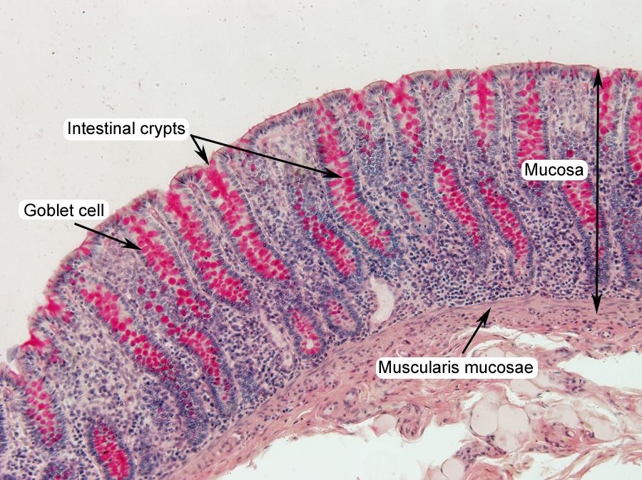
Colon diagram with labels. The Colon - Ascending - Transverse - Descending - Sigmoid ... The colon (large intestine) is the distal part of the gastrointestinal tract, extending from the cecum to the anal canal. It receives digested food from the small intestine, from which it absorbs water and electrolytes to form faeces. Anatomically, the colon can be divided into four parts - ascending, transverse, descending and sigmoid. Histology | Colon This diagram illustrates the 4 basic layers of the colon. The inner pink layer is the mucosa, the yellow layer beneath the mucosa is called the submucosa, while the red layer is the muscular layer (muscularis) and the 4 th layer is called the serosa or adventitia. Courtesy Ashley Davidoff MD 32338 Ultrasound of normal large bowel Label Digestive System Diagram Printout ... Read the definitions below, then label the digestive system anatomy diagram. anus - the opening at the end of the digestive system from which feces (waste) exits the body. appendix - a small sac located on the cecum. ascending colon - the part of the large intestine that run upwards; it is located after the cecum. Anatomy of Colon and Rectum | SEER Training Anatomy of Colon and Rectum. The entire colon is about 5 feet (150 cm) long, and is divided into five major segments. The rectum is the last anatomic segment before the anus.. The ascending and descending colon are supported by peritoneal folds called mesentery.. The right colon consists of the cecum, ascending colon, hepatic flexure and the right half of the transverse colon.
How does the bowel work? Bowel information with diagrams ... The large bowel is made up of the colon, rectum and anus. Diagram of the digestive system. When you swallow food, it passes down the gullet (oesophagus) to the stomach. This is where digestion begins. The food then enters the small bowel, where nutrients and minerals from food are absorbed. The digested food then moves into the colon. Colon Picture Labeling Flashcards - Quizlet Colon Picture Labeling STUDY Flashcards Learn Write Spell Test PLAY Match Gravity Created by XertziePLUS Terms in this set (11) rectum 10 external anal sphincter 12 teniae coli 14 cecum 8 vermiform appendix 9 transverse colon 2 ascending colon 5 ileum 6 ileocecal valve 7 sigmoid colon 13 descending colon 16 YOU MIGHT ALSO LIKE... Illustration Picture of Anatomy - Colon The colon is the largest part of the large intestine, extending from the cecum to the rectum. It is 5 feet long and its function is to reabsorb water from digested food and concentrate solid waste material, known as stool. The colon is made of several sections. The ascending colon travels up the right side of the abdomen. Digestive tract - lower GI tract labeled - labeled | Media ... Illustration of the digestive tract within an outline of the top half of a human body. The appendix, cecum, colon, sigmoid colon, rectum, stomach, large intestine, small intestine, and anus are labeled. Alternate Text Illustration of the digestive tract within an outline of the top half of a human body.
Colon Anatomy (with Small Intestine Label): Image Details ... 720x602. View. Download. Title: Colon Anatomy (with Small Intestine Label) Description: Drawing shows the cecum, ascending colon, transverse colon, descending colon, sigmoid colon, rectum, and anal canal. Also shown is the small intestine. The cecum connects the small intestine to the colon. Sigmoid colon - Definition, Anatomy and Function | Kenhub Sigmoid colon Author: Shahab Shahid MBBS • Reviewer: Jerome Goffin Last reviewed: October 04, 2021 Reading time: 8 minutes The sigmoid colon is part of the hindgut.It is the last part of the colon before the rectum.It acts as a site for water absorption from the faeces, as a site for flatus to be stored before being expelled, and as a site of peristalsis. 40 Colon diagram Vector Images, Colon diagram ... 40 Colon diagram Stock Vector Images, Royalty-free Colon diagram Drawings & Illustrations. VectorMine Crohns disease vector illustration. Labeled diagram with diagnosis. VectorMine Ulcerative colitis vector illustration. Labeled anatomical infographic. switch…case in C (Switch Statement in C) with Examples Apr 09, 2022 · Case labels must be constants and unique. Case labels must end with a colon ( : ). A break keyword must be present in each case. There can be only one default label. We can nest multiple switch statements. Summary. A switch is a decision making construct in ‘C.’ A switch is used in a program where multiple decisions are involved.
According2Robyn: Digestive System, Part 9: The Colon
Pascal Case Statement - Tutorialspoint You can have any number of case statements within a case. Each case is followed by the value to be compared to and a colon. The case label for a case must be the same data type as the expression in the case statement, and it must be a constant or a literal. The compiler will evaluate the case expression.

OB Images
Intestines (Anatomy): Picture, Function, Location, Conditions The large intestine (colon or large bowel) is about 5 feet long and about 3 inches in diameter. The colon absorbs water from wastes, creating stool. As stool enters the rectum, nerves there create ...

Abdomen Picture Image on MedicineNet.com
Abdomen and digestive system anatomy: diagrams labeled Full labeled anatomical diagrams - Anatomy of the abdomen and digestive system: these general diagrams show the digestive system, with the major human anatomical structures labeled (mouth, tongue, oral cavity, teeth, buccal glands, throat, pharynx, oesophagus, stomach, small intestine, large intestine, liver, gall bladder and pancreas).
Digestive
PDF ANATOMIC DRAWINGS OF THE DIGESTIVE SYSTEM Esophageal ... Colon (C18._).0 Cecum.1 Appendix.2 Ascending.3 Hepatic flex..4 Transverse.5 Splenic flex..6 Descending.7 Sigmoid.8 Overlapping.9 Colon, NOS Yes Yes Yes Yes Yes Yes Yes Yes Yes Yes Yes Yes Yes Yes Yes Yes Yes Yes Yes Yes Yes Yes Yes Yes Yes Yes Yes Yes Yes Yes Yes Yes Yes Yes Yes Yes Yes Yes Yes Yes Yes Yes Yes Yes Yes Yes Yes Yes Yes Yes Yes ...

Gastrointestinal System
Small Intestine and Large Intestine (With Diagram) 2. Propulsion: The chyme is moved over the large area of small intestine to facilitate digestion and absorption and the residues are propelled downwards to the ileocecal junction to reach the large intestine, mostly for excretion. In man, the time taken for the food to travel in the small intestine as well as in stomach can be easily estimated ...

Colon Diagram - ColoCleanz
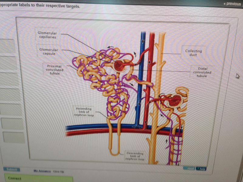
Labeled Diagram of the Human Kidney - Bodytomy The renal medulla comprises a set of 8-18 conical structures called renal pyramids that are surrounded by the cortex. Portions of the cortex between two adjacent pyramids are termed as renal columns. Spread in these pyramids and the cortex, are the functional units callednephrons. The actual filtration of blood occurs in the nephrons.

The Colon
PDF Digestive System Diagram Label these: #1 (Urine/pee pee) #2 (Solid Waste/poop) Hydrochloric Mechanical Digestion Chemical Digestion Saliva Acid Pepsin Bile Lipase Stomach Small Intestine Enzymes from Liver and Pancreas Large Intestine (Transverse Colon) Descending ColonCirculatory System Kidneys #1 #2 Water and Vitamins Nutrients The Digestive System
Patient Resources | Gastroenterology and Hepatology
Colon Diagram Stock Illustrations - 3,131 Colon Diagram ... Labeled Diagram. Human colon. Colon - lymphatic drainage. Pathways of lymphatic drainage of the colon. Anatomy of the Colon, Rectum and Anus. Digestive system, large intestine lymphatic. Colonoscopy procedure labeled 3d diagram on white background. Eps 10. Crohns disease vector illustration. Labeled diagram with diagnosis. Crohns disease vector ...

Large Intestine Histology - Colon (labels) - histology slide - | Histology slides, Tissue ...
Large Intestine Anatomy, Parts, Diagram & Major Function ... Discuss the role of the large intestine in humans, the anatomy, and the function of the large intestine. Draw a diagram of the large intestine and label all parts.
Anatomy and physiology of the colon | General center | SteadyHealth.com
Digestive System Labeled - Agaliprogram digestive system labeled. #1 (urine/pee pee) #2 (solid waste/poop) hydrochloric mechanical digestion chemical digestion saliva acid pepsin bile lipase stomach small intestine enzymes from liver and pancreas large intestine (transverse colon) descending coloncirculatory system there is an unlabeled diagram in the end of the article for readers to …
How The Colon Works | Colon Cleanse Recipes | Homemade Colon Cleanse
Colon: Anatomy, histology, composition, function | Kenhub The colon forms part of the large intestine and extends between the caecum and the rectum. It is about 1.5 meters in length and consists of four parts: ascending transverse descending sigmoid colon You can recognize it easily through several distinct morphological features like semilunar folds and pouches called haustra.
Chapter 25: The Urinary System (Mastering) Flashcards | Easy Notecards
the large intestine labeling diagram Diagram | Quizlet Start studying the large intestine labeling diagram. Learn vocabulary, terms, and more with flashcards, games, and other study tools.

Colon | Obgyn Key
Colon Anatomy - Human Body Diagrams - Medical Art Library The large intestine is divided into the cecum, colon, rectum and anal canal.The large intestine begins at the cecum. The ileum (small intestine) ends where it connects to the cecum at the ileocecal junction.. The colon is divided into four parts: the ascending, transverse, descending and sigmoid.The ascending and transverse colon meet at the right hepatic flexure (near the liver).
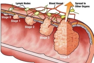
colon cancer stage | Anatomy System - Human Body Anatomy diagram and chart images
Colonoscopy Measurements (cm) from Anal Verge | SEER Training Home » Site-specific Modules » Colorectal Cancer » Anatomy of Colon and Rectum » Colonoscopy Measurements (cm) from Anal Verge Section Menu Cancer Registration & Surveillance Modules
Post a Comment for "41 colon diagram with labels"