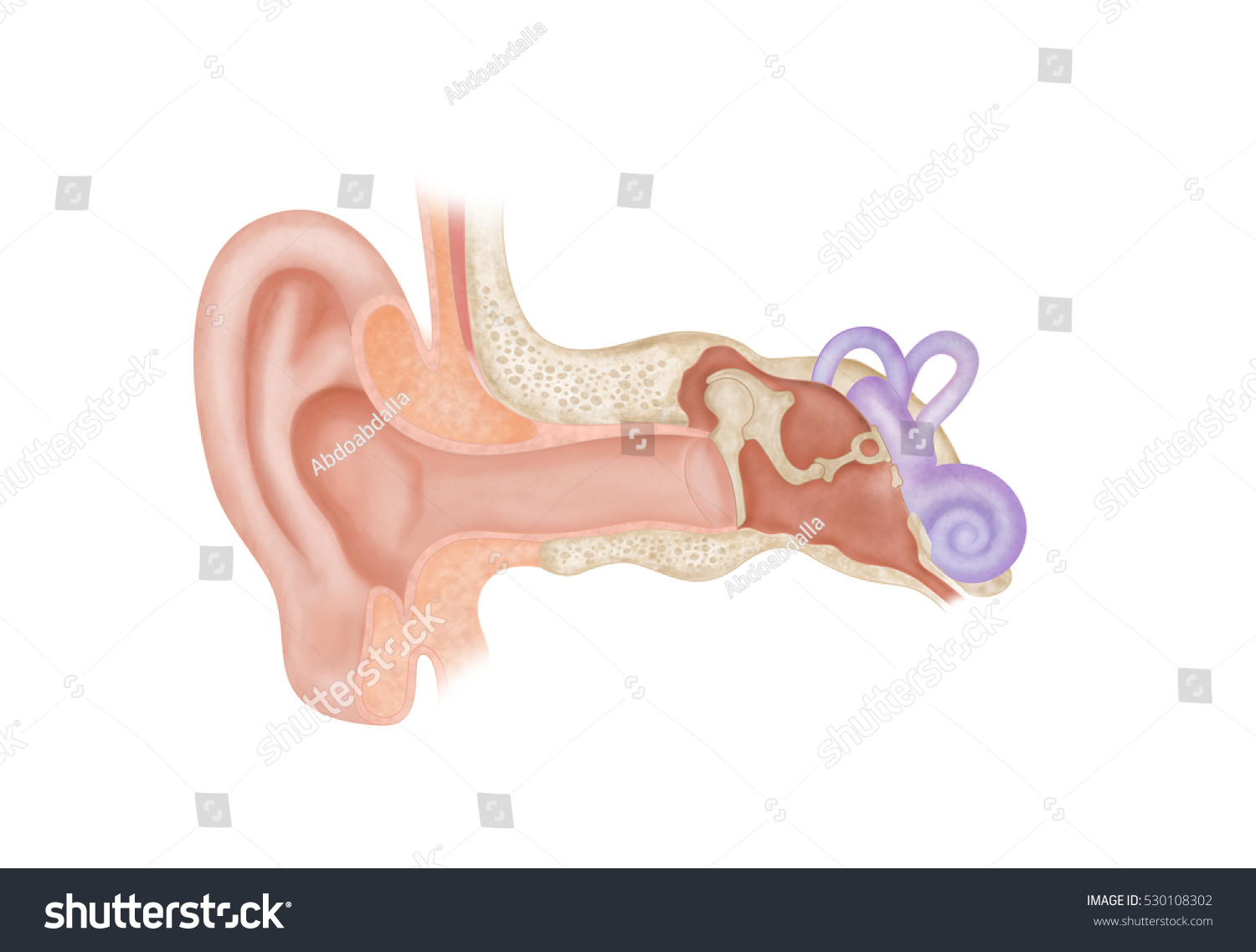
43 ear anatomy without labels
› enAnatomy, medical imaging and e-learning for healthcare ... IMAIOS and selected third parties, use cookies or similar technologies, in particular for audience measurement. Cookies allow us to analyze and store information such as the characteristics of your device as well as certain personal data (e.g., IP addresses, navigation, usage or geolocation data, unique identifiers). › publication › 344058244FUNDAMENTAL PRINCIPLES OF HUMAN ANATOMY & PHYSIOLOGY Jul 02, 2016 · Labels of human body features displayed on images of actual human bodies, from which body hair and male facial hair has been removed. (Source: City Studios in Stockholm ( ...
Label Anatomy Dogs Teaching Resources | Teachers Pay Teachers 3.0. (1) $2.50. PDF. Label the Parts of a DogThis is a perfect addition to your Chinese New-year Lessons. Printables - 12 parts• Colour Printout - A control chart for independent work• Printout 1- boxes with dashes for cutting and gluing• Printout 2- with the words at the bottom, for writing Other Dog Lessons:2018 Year of the Dog ~ Dog ...

Ear anatomy without labels
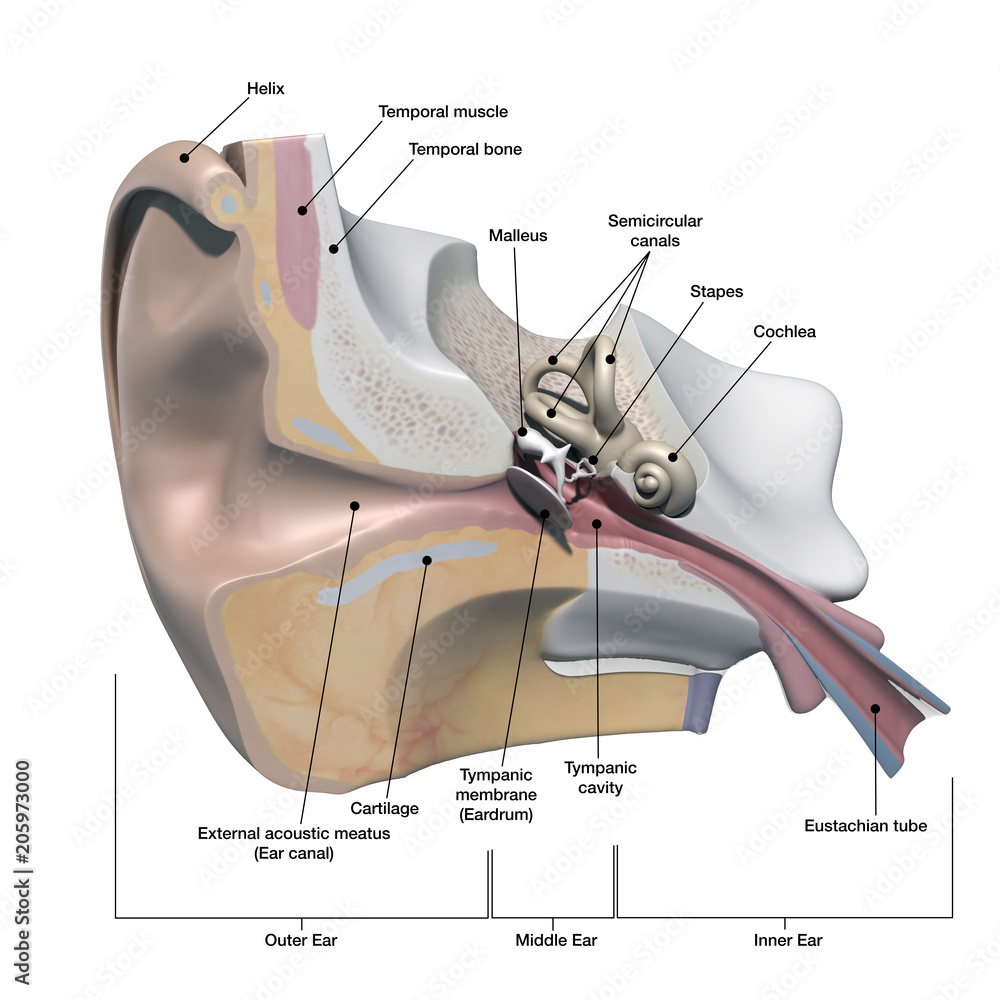
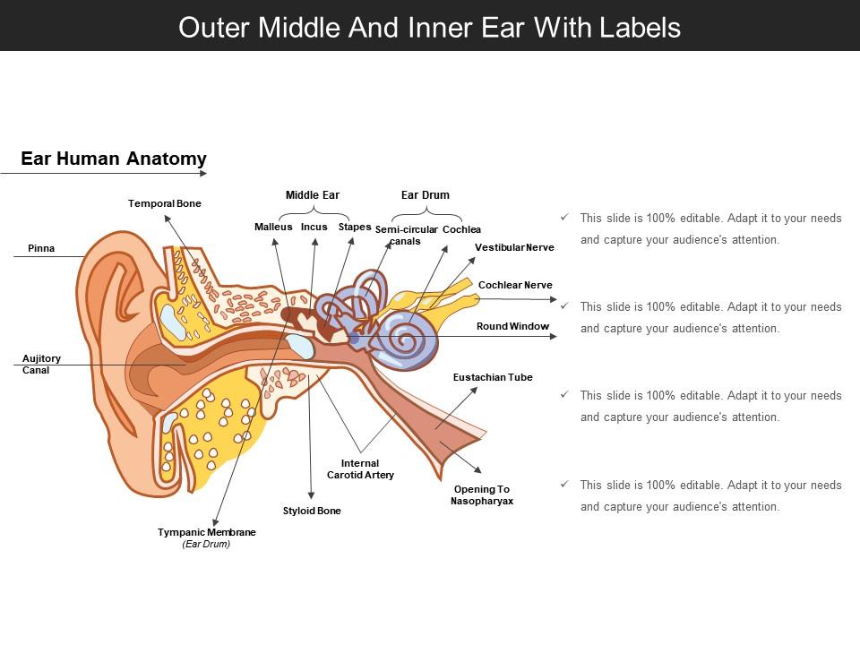
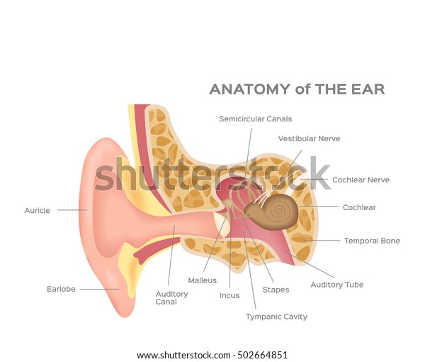
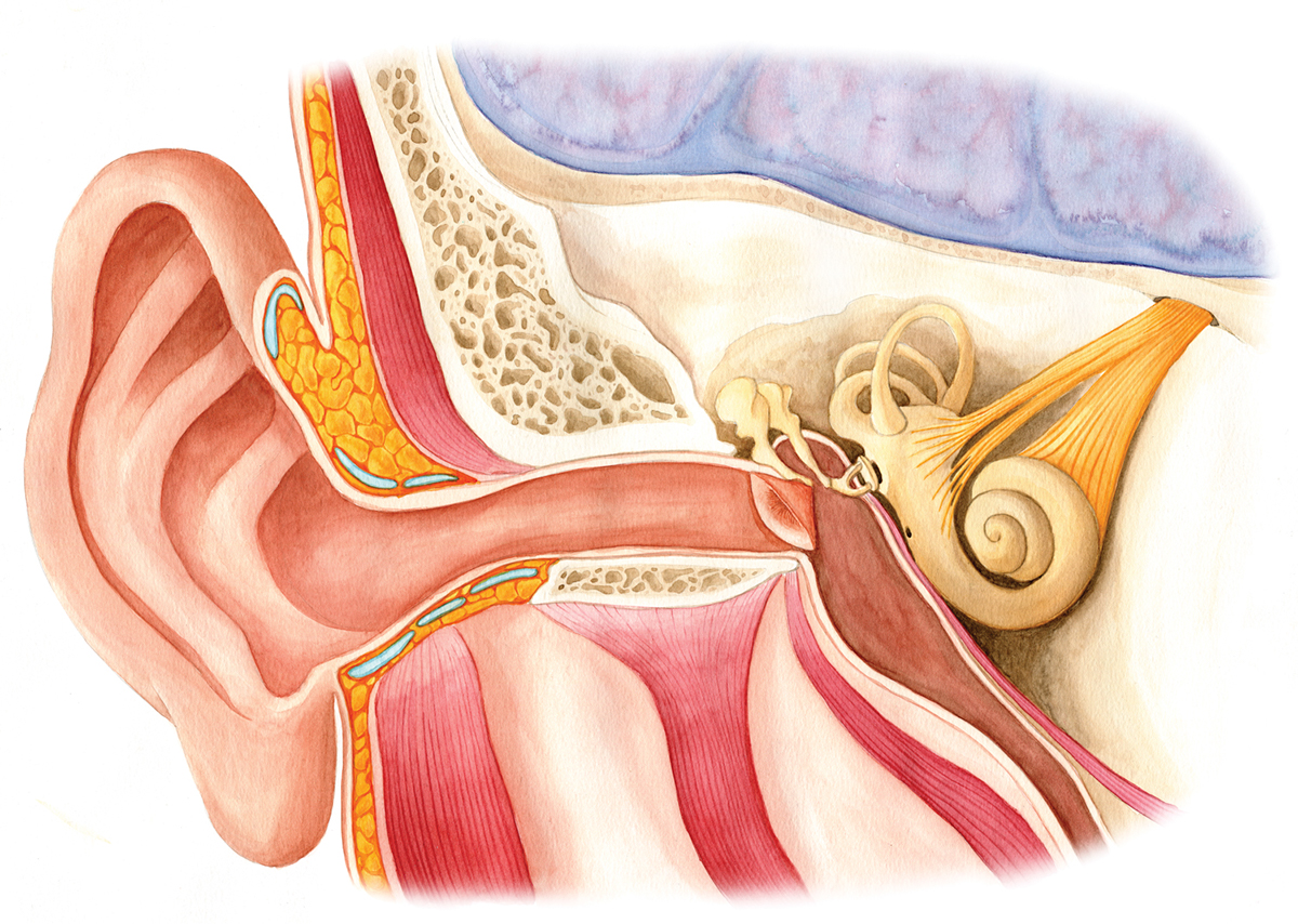
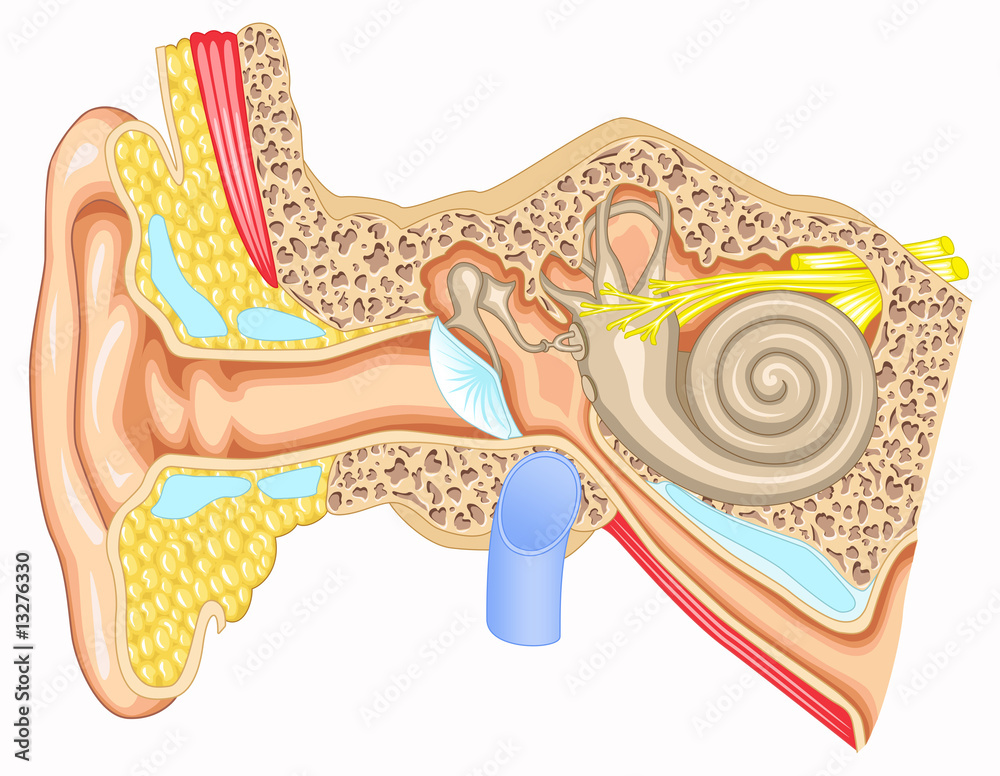
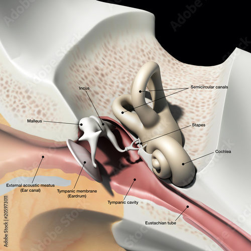
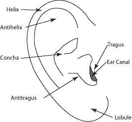
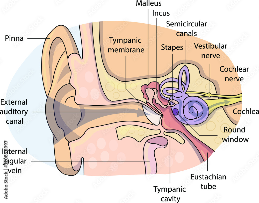
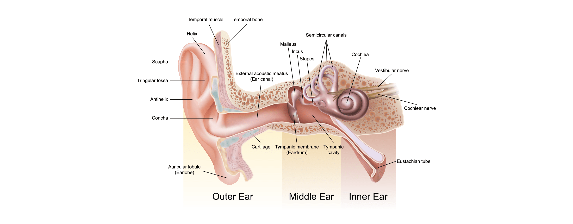
Ear Anatomy - Outer Ear | McGovern Medical School The medical term for the outer ear is the auricle or pinna. The outer ear is made up of cartilage and skin. There are three different parts to the outer ear; the tragus, helix and the lobule. EAR CANAL The ear canal starts at the outer ear and ends at the ear drum. The canal is approximately an inch in length. Image result for ear structure without label | Ear anatomy, Human ear ... Each nephron is made of two main parts called malpighian body and covoluted tubule.Malphigian body is double layered cup also called bowman`s capsule. The inner cup consists network of capillaries called Glomerulus. Now let`s start the diagram. 1.Draw a egg shape and make a folded curve as shown in the figure and… L Amy Yee Anatomy Well-Labelled Diagram Of Ear With Explanation - BYJUS Eustachian Tube is a tube that connects the middle ear to the back of the nose. It helps to maintain equal pressure in the middle ear which facilitates the proper transmission of sound waves. The Inner ear consists of Cochlea that comprises the nerves of hearing. Semicircular canals contain the receptors that help in maintaining balance.
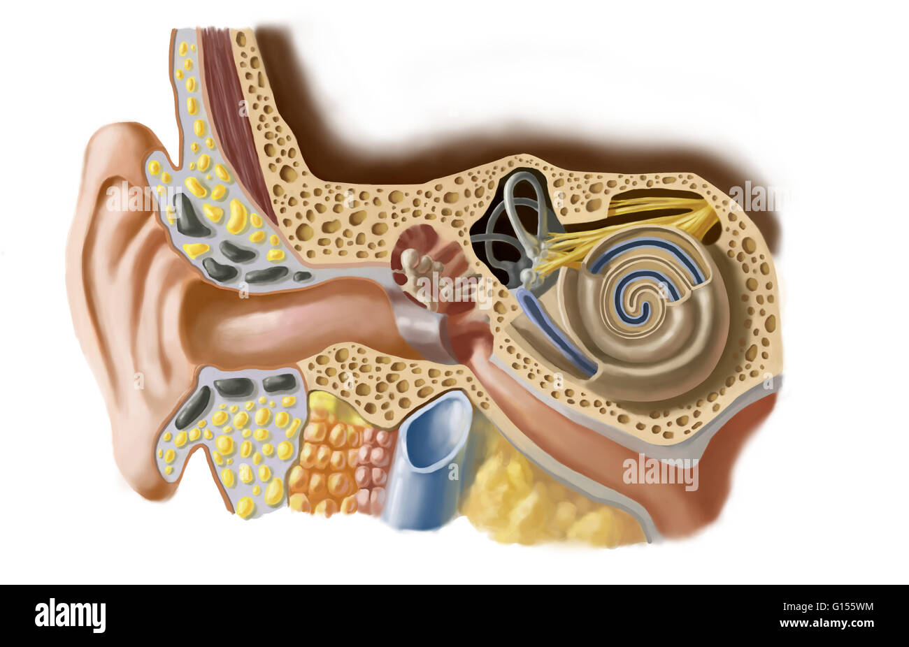
Ear anatomy without labels. Human Ear: Structure and Anatomy - Online Biology Notes Ear ossicles: The three ear ossicles (malleus, incus and stapes) form a chain of lever extending from tympanic membrane to inner ear. The ear ossicles transmit sound wave from ear drum to inner ear. Ear ossicles communicate the ear drum with internal ear through fenestra ovalis ( oval window). The ear ossicles are; Human Ear Diagram - Bodytomy Auditory Ossicles: The three small bones in the middle ear, called malleus, stapes, and incus, are connected. These bones together are called the auditory ossicles, and their purpose is to let the sound that strikes the eardrum, further into the inner ear. Ear Anatomy Images | McGovern Medical School Ear Anatomy Images. The ear drum is often transparent and looks like a stretched piece of clear plastic. The drum is approximately the size of a dime, with the newborn ear drum the same size as the adult. The malleus is the middle ear bone which is attached to the drum and easily identified. The middle ear space can be seen through the ear drum ... Picture of the Ear: Ear Conditions and Treatments - WebMD Earache: Pain in the ear can have many causes. Some of these are serious, some are not serious. Otitis media (middle ear inflammation): Inflammation or infection of the middle ear (behind the ...
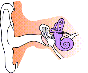
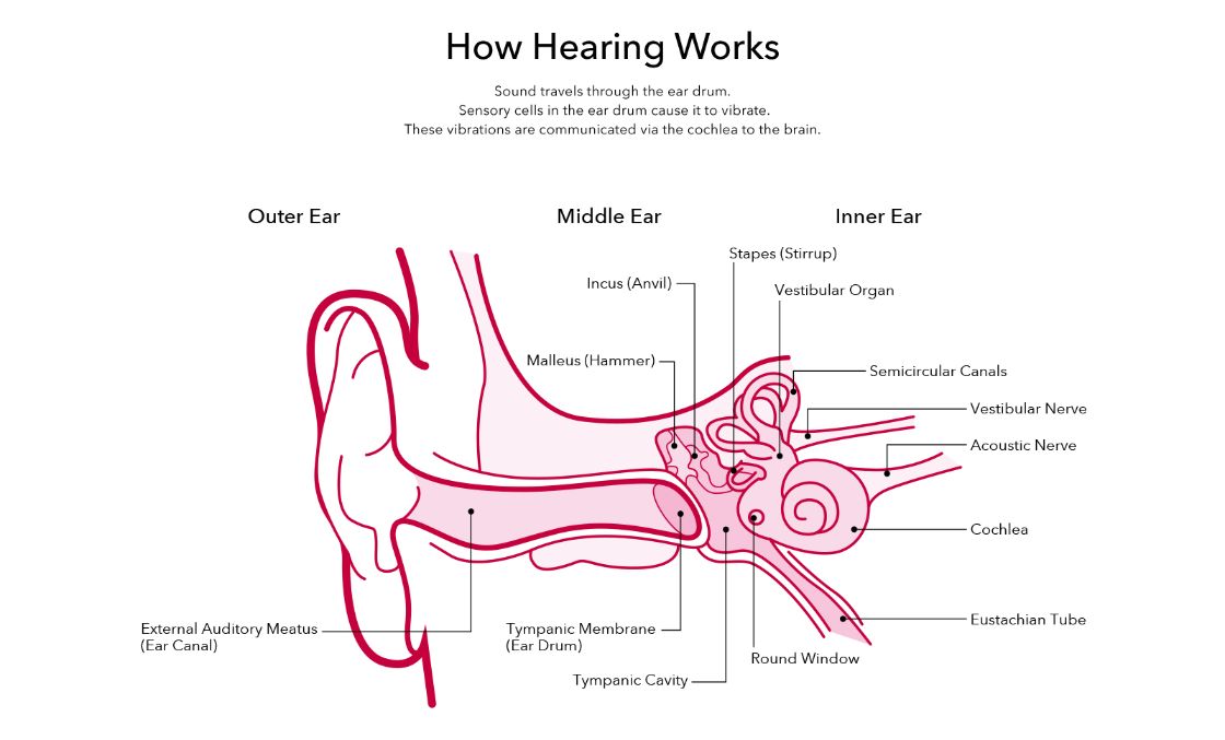
Ear Diagram and Labeling Worksheet / Worksheet - Twinkl This allows you to tailor the task to the individual abilities of your learners. The first worksheet presents an ear with annotations showing the first letters of its key features. For example, a label marked 'P' links to the Pinna (outer ear). The second page shows an ear diagram without labels. The final page shows the labels linking to the ... › en › e-AnatomyNormal chest MDCT with anatomic labels | e-Anatomy - IMAIOS Mar 10, 2022 · IMAIOS and selected third parties, use cookies or similar technologies, in particular for audience measurement. Cookies allow us to analyze and store information such as the characteristics of your device as well as certain personal data (e.g., IP addresses, navigation, usage or geolocation data, unique identifiers). Image result for ear structure without label - Pinterest Preschool Kid Learning Ear Coloring Pages to Color, Print and Download for Free along with bunch of favorite Ear coloring page for kids. Simply do online coloring for Preschool Kid Learning Ear Coloring Pages directly from your gadget, support for iPad, android tab or using our web feature. D. Darina Lețu. Doodle and Art. Human Ear Anatomy - Parts of Ear Structure, Diagram and Ear Problems The external (outer) ear consists of the auricle, external auditory canal, and eardrum (Figure 1 and 2). The auricle or pinna is a flap of elastic cartilage shaped like the flared end of a trumpet and covered by skin. The rim of the auricle is the helix; the inferior portion is the lobule. Ligaments and muscles attach the auricle to the head.
Ear Labels Flashcards | Quizlet Terms in this set (16) auricle external auditory canal tympanic membrane malleus (hammer) Incus (anvil) stapes (stirrup) auditory/eustachian/pharyngotympanic tube vestibules semicicular canals Ampulla of semicircular canals round window oval window and round window cochlea snail cochlear duct in cochlea vestibular nerve Parts of the Ear Labelled Diagram Activity | Twinkl The first worksheet presents an ear with annotations showing the first letters of its key features. For example, a label marked 'P' links to the Pinna (outer ear). The second page shows an ear diagram without labels. The final page shows the labels linking to the beginning letters of each feature, but without the words list. › en › libraryAnatomy coloring books: How to use & free PDF | Kenhub Sep 14, 2022 · Generally, an anatomy coloring book will divide subject matter into sections, with each section containing many topics. For each topic you will find black and white anatomical drawings, often accompanied by labels, related text and terminology. Tired of keeping track of so many study materials? The Ear: Anatomy, Function, and Treatment - Verywell Health The middle ear (also known as the tympanum or tympanic cavity) is a complicated network of tunnels, chambers, openings, and canals mostly inside openings within the temporal bone on each side of the skull. The 2 largest chambers are called the middle ear space and mastoid.
Inner ear - Wikipedia
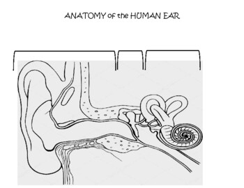
label the ear worksheet 14 Best Images Of Ear Hearing Worksheets - Listening Ear Craft Template ear worksheet diagram inner anatomy answers parts eye animals physiology labelled worksheets ossicles senses auditory middle hearing outer pinna canal Ear Diagram Without Labels & With Them - Labelling Worksheet
Human Ear Anatomy Cross Section View with Labels on White ...
255 Human Ear Diagram Premium High Res Photos - Getty Images external auditory canal of human ear (with labels). - human ear diagram stock illustrations engraved antique, anatomy of the ear and nose engraving antique illustration, published 1851 - human ear diagram stock illustrations
Ear diagram - Teaching resources
appgrooves.com › rank › medicalBest 10 Anatomy Apps - Last Updated July 26, 2022 - AppGrooves May 01, 2018 · This app is absolutely phenomenal. I have used it for 6 years now. The fact that you are able to explore in zoomable detail the thousands of muscles, tendons, ligaments joints, bones, organs, attachment points, connections, surfaces, etc., has been so enlightening for me in exploring the anatomy involved in the random injuries I have passed in life, including injuries that I never knew of ...

Anatomy Of The Human Ear Blank - Ear Anatomy, HD Png Download ...
eye and ear anatomy worksheet eye diagram brain blank printable anatomy label science without structure clipart cliparts class activities library experiments rudyard fair Label the ear worksheets (sb6635). Ear psychology sensation auditory ears human dog general process biology working cleaning diagram anatomy dogs project bionic hearing source tos.
Anatomy of the Ear Parts of the Ear Minimum time needed 12 ...
Enchanted Learning Moved Permanently. The document has moved here.
Outer Middle And Inner Ear With Labels | PowerPoint ...
Ear (Anatomy): Overview, Parts and Functions | Biology Dictionary The ear canal is the opening through which sound waves enter the middle ear. It serves to further focus and concentrate the vibrations collected by the pinna, ensuring that the vibrations will be clear and strong enough to be amplified and turned into nerve impulses. The ear canal is only 2-3 centimeters deep - a little bit less than one inch.

Label a diagram that includes the outer, middle, and inner ...
Outer Ear: Anatomy, Location, and Function - Verywell Health Fossa, superior crus, inferior crus, and antihelix: These sections make up the middle ridges and depressions of the outer ear. The superior crus is the first ridge that emerges moving in from the helix. The inferior crus is an extension of the superior crus, branching off toward the head. The antihelix is the lowest extension of this ridge.

The Human Ear — Anatomy and Function - Divers Alert Network
Label Parts of the Human Ear - University of Dayton Parts of the Ear. Select the correct label for each part of the ear. Click on the Score button to see how you did. Incorrect answers will be marked in red.
Solved] Use the word list on the following page to label the ...
Blank ear diagrams and quizzes: The fastest way to learn ear anatomy Ear diagrams (labeled and unlabeled) Accelerate your learning with interactive quizzes Sources + Show all Ear anatomy overview Although it's not obvious to look at, the ear is anatomically divided into three portions: External (outer) ear Middle ear Inner ear As you can imagine, there's a lot of associated anatomy to learn for each portion!
Human Ear Structure Medical Educational Science Stock Vector ...
14,026 Human ear anatomy Images, Stock Photos & Vectors - Shutterstock 14,026 human ear anatomy stock photos, vectors, and illustrations are available royalty-free. See human ear anatomy stock video clips Image type Orientation Color People Artists Sort by Popular Healthcare and Medical Anatomy ear cochlea medicine inner ear organ human body biology diagram Next of 141
Ear Anatomy Without Labels Digital Art Stock Illustration ...
Image result for ear structure without label | Ear diagram, Human ear ... Feb 12, 2018 - Image result for ear structure without label. Feb 12, 2018 - Image result for ear structure without label. Pinterest. Today. Explore. When the auto-complete results are available, use the up and down arrows to review and Enter to select. Touch device users can explore by touch or with swipe gestures.

Anatomy of the nose and throat. Human organ structure ...
Anatomy of the Ear | Geeky Medics The tympanic membrane, or eardrum, marks the border between the external and middle ear. It is formed of a middle layer of connective tissue with a layer of skin on its lateral surface (facing the external acoustic meatus) and mucous membrane on its medial surface (facing the middle ear).
Anatomy of the Human Ear: Identify, Label and Color
Anatomical terms: How to memorize them in 4 steps | Kenhub Create a study schedule. Anatomy is another language, so learning its words takes time. Divide them evenly and learn or review some of them each day. Visualize the concepts. Don't just stare at an atlas - take the structure that you're looking at and visualize it in your head. Memorize efficiently. Link the word to the structure by creating a ...
Auditory pathway: Anatomy, ear structures, transduction | Kenhub
Anatomy of the Ear | Inner Ear | Middle Ear | Outer Ear The Middle Ear. The middle ear includes: eardrum. cavity (also called the tympanic cavity) ossicles (3 tiny bones that are attached) malleus (or hammer) - long handle attached to the eardrum. incus (or anvil) - the bridge bone between the malleus and the stapes. stapes (or stirrup) - the footplate; the smallest bone in the body.
File:Ear-anatomy-notext-small.png - Wikimedia Commons
quizlet.com › 414265947 › anatomy-lecture-midtermAnatomy Lecture Midterm Flashcards | Quizlet Study with Quizlet and memorize flashcards containing terms like Place a single word into each sentence to make it correct. Then rearrange the sentences into the correct order to explain the process of the cardiocyte action potential. +30mV Chloride Ion Negative Positive -55mV Resting Calcium ion Cardiomyocyte Once these channels close, potassium ions flow out quickly and restore the ...

Labelling the EAR --- This is a one page worksheet that ...
Human Middle Ear Anatomy Cross Section View With Labels Stock Photo ... Description Computer generated image of the human middle ear bones and inner ear with anatomical labeling. 1 credit Essentials collection for this image $4 with a 1-month subscription (10 Essentials images for $40) Continue with purchase View plans and pricing Includes our standard license. Add an extended license. Credit: Hank Grebe

The ear canal: Anatomy, diagram, and common conditions
Ear Anatomy without Labels, Digital Art - Shutterstock Ear Anatomy Without Labels Digital Art Stock Illustration 530108302 Download for free See more Popularity score High Usage score High usage Superstar Shutterstock customers love this asset! Item ID: 530108302 Ear Anatomy without Labels, Digital Art Formats 8976 × 6201 pixels • 29.9 × 20.7 in • DPI 300 • JPG
Ear Anatomy for Visual Guide to ENT Pathology on Behance
ecorche.anatomy4sculptors.com › head-and-neck › earEcorche Reference Tool | Anatomy For Sculptors Ecorche Reference Tool has 3D references of the whole human figure. The 3D models feature muscle motion, bony landmarks, facial muscles, block-outs, etc.
Ear anatomy - Cross section view Stock Illustration | Adobe Stock
Well-Labelled Diagram Of Ear With Explanation - BYJUS Eustachian Tube is a tube that connects the middle ear to the back of the nose. It helps to maintain equal pressure in the middle ear which facilitates the proper transmission of sound waves. The Inner ear consists of Cochlea that comprises the nerves of hearing. Semicircular canals contain the receptors that help in maintaining balance.
Ear Anatomy | How Does The Ear Work? | Amplifon AU
Image result for ear structure without label | Ear anatomy, Human ear ... Each nephron is made of two main parts called malpighian body and covoluted tubule.Malphigian body is double layered cup also called bowman`s capsule. The inner cup consists network of capillaries called Glomerulus. Now let`s start the diagram. 1.Draw a egg shape and make a folded curve as shown in the figure and… L Amy Yee Anatomy
Human Ear Anatomy Detailed View with Labeling Stock ...
Ear Anatomy - Outer Ear | McGovern Medical School The medical term for the outer ear is the auricle or pinna. The outer ear is made up of cartilage and skin. There are three different parts to the outer ear; the tragus, helix and the lobule. EAR CANAL The ear canal starts at the outer ear and ends at the ear drum. The canal is approximately an inch in length.
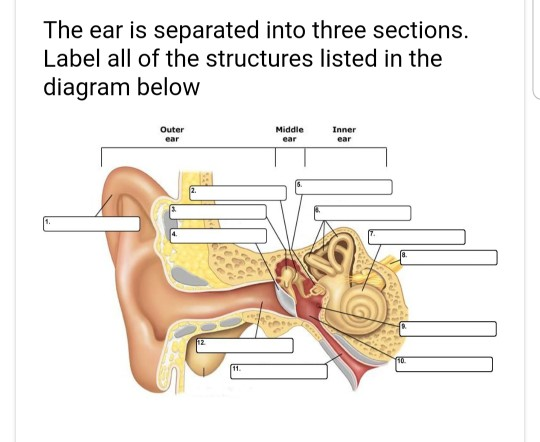
Solved The ear is separated into three sections. Label all ...

Anatomy of the Inner Ear | Doctor Stock

ch 10: ear labeling Diagram | Quizlet

Ear labeling Diagram | Quizlet
Ear Anatomy – Outer Ear | McGovern Medical School
Ear Anatomy – Drag and Drop Activity
Ear diagram hi-res stock photography and images - Alamy

The structure and function of the ear and its role in hearing ...

5,905 Ear Anatomy Stock Photos, Pictures & Royalty-Free ...

Structures and functions of Ears Diagram | Quizlet
2,107 Inner Ear Illustrations & Clip Art - iStock

ear labeling Diagram | Quizlet

1: Diagram showing the structure of the human ear, detailing ...
Vector illustration of a schematically painting of an ear ...
The Anatomy of the Hearing System – Marin Hearing Center

Cochlea Implants for the Deaf and Severely Hard of Hearing ...
Ear anatomy: Parts and functions | Kenhub

Image result for ear structure without label | Ear diagram ...

Label the ear structures. | Homework.Study.com
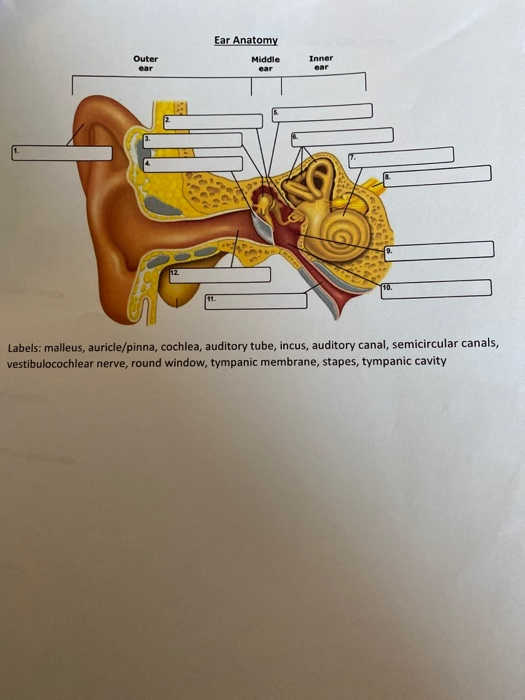
Solved Ear Anatomy Middle Outer Inner 1. Labels: malleus ...

Child Ear Model | Human Body Anatomy Replica of Child's Ear ...

anatomy module 4 lab exam - label the ear model Diagram | Quizlet
Post a Comment for "43 ear anatomy without labels"